Sight is a sense faculty to see people and things around us. Visual Acuity defines the clarity of the visual perception of an image. Vision can be lost at birth or during one’s lifetime, painlessly or painfully for various reasons.
Pain in vision draws one’s attention to seek medical treatment. Painless loss of vision however is akin to termites silently causing destruction from the inside without initial symptoms. One common condition which causes permanent loss of vision is Glaucoma.
When does GLAUCOMA occur?
Glaucoma is a condition where the optic nerve is damaged from increased intraocular pressure without us being aware. Glaucoma damage is permanent and it is not reversible!
Aqueous humor controls the pressure inside the eyes to keep the eyeball firm. It is secreted in the ciliary body and passed through the pupil into the space between the iris and the cornea. Aqueous is drained through the Trabecular meshwork at the anterior chamber angle (Fig 1).
There are many risk factors associated with gloucoma and they include:
- Being over 40 years old
- Having east-Asian/African American ancestry
- Family history of glaucoma
- People who have high eye pressure
- Being far-sighted or near-sighted
- Having an eye injury
- Taking steroid via eye drops or orally
- Having a thinner central corneal thickness
- Having a thinning optic nerve
- Being diabetic
- Having constant migraines
- Having hypertension
- Having other health-related problems
Glaucoma has no symptoms in early stages. Many people are unaware of this silent but preventable blindness. Regular eye screening helps to detect this disease before vision loss.
World Health Organization Eye Screening Recommendation
Glaucoma Screening for normal individuals:
- Once in every 2 years for persons 40 years and above
- Yearly for persons 50 years and above
- Screen for intraocular pressure if immediate family is diagnosed with glaucoma
American Academy of Ophthalmology Recommendation
- Full eye examination every 5 to 10 years if a person is below 40 years and has no known risk factors for Glaucoma.
- Screening schedules for individuals at risk of glaucoma:
1 – 3 yearly for 40 – 50 years old
1 – 2 yearly for 55 – 64 years old
6 – 12 monthly for 65 years and above
How is Glaucoma treated?
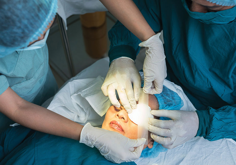
Prognosis through medication and surgery to prevent further optic nerve damage is the best way.

Treatment options include:
- Glaucoma eye drop medications. Used daily, the eye drops lower intraocular pressure by reducing the amount of aqueous fluid produced by the eyes or helps the fluid flow better through the drainage angle.
- Surgery to help drain the aqueous fluid from the eye.
The various procedure types can be carried out in the ophthalmologist’s clinic or as an outpatient in the surgical centre:
1. Trabeculoplasty
-
- This surgery is for persons with open-angle glaucoma.
- The surgeon uses the laser to facilitate the drainage angle function better, in order to reduce the eye pressure.
2. Iridotomy
-
- This procedure is recommended for persons with angle-closure glaucoma. The ophthalmologist uses the laser to create a tiny hole in the iris allowing the fluid to flow towards the drainage angle.
3. Trabeculectomy
-
- The surgeon creates a tiny flap in the sclera and a bubble in the conjunctiva called a filtration bleb to drain the aqueous humor (Fig 2).
- In the bleb, the fluid is absorbed by the tissue around the eye, hence lowering the eye pressure.
4. Glaucoma Drainage Implant
-
- The ophthalmologist may implant a tiny drainage tube in the eye (Fig 3).
- It sends the fluid to a collection area called a reservoir.
- The surgeon creates the reservoir beneath the conjunctiva.
- The fluid is absorbed into nearby blood vessels.
Keep in mind
- Team effort between patients and doctor
- Adhere to doctor’s instructions
- Visit the doctor every 3 – 6 months
- Refer enquiries to consultant
 Dr. Mohd Hassan @ Maung Maung Win
Consultant Ophthalmologist,
Columbia Asia Hospital – Setapak


